THE FULL JOURNEY
From a single slice
to the whole brain
Start from one black-and-white MRI slice and work up through the region map, the 3D atlas, and comparisons of aging and disease — five chapters, one step at a time.
01
CHAPTER 01 · Meeting your first MRI slice
Black-and-white MRI, scrolled with your fingertip
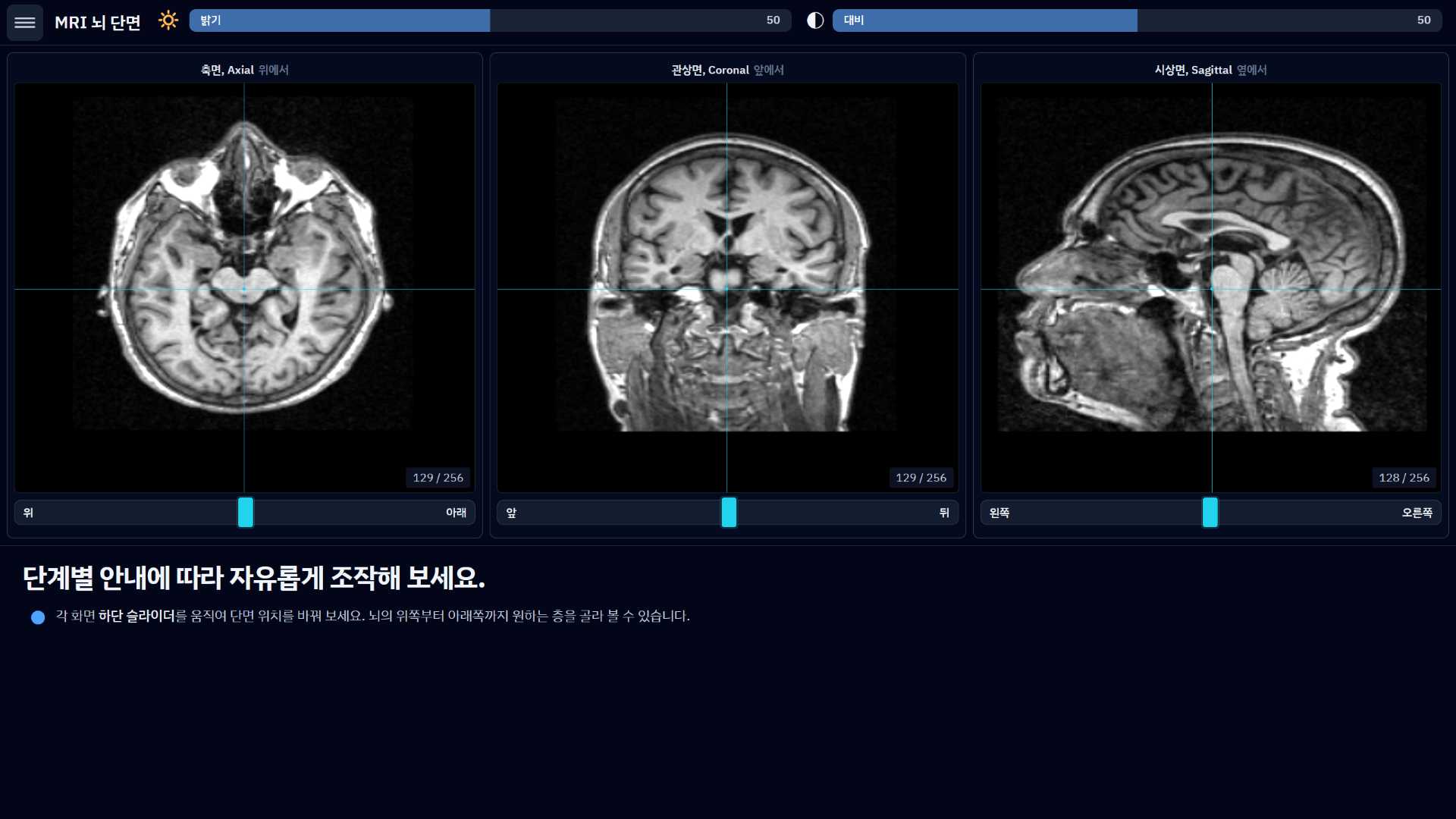
Brain scans aren't as hard as they look. Scroll through the same black-and-white MRI a hospital takes, right at your fingertip. Adjust brightness and contrast, look inside the brain from three directions, and watch the moment color lands on all that complicated structure.
01 / 05
02
CHAPTER 02 · The region map
Point at a spot and only that region stays sharp in the 3D brain
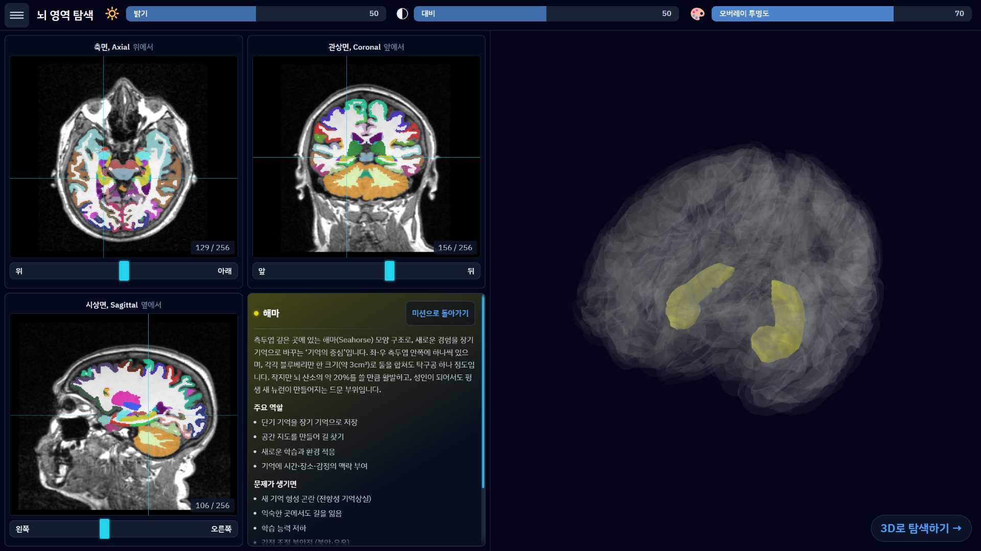
What does this part actually do? Point at a spot on the slice and everything else in the 3D brain fades back — only that region stays sharp. Turn the view around and pick up, naturally, where memory, emotion and language live.
02 / 05
03
CHAPTER 03 · The 3D atlas
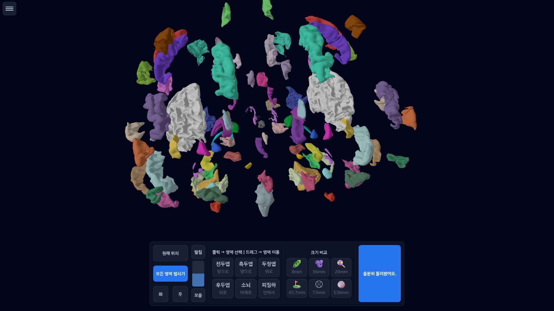
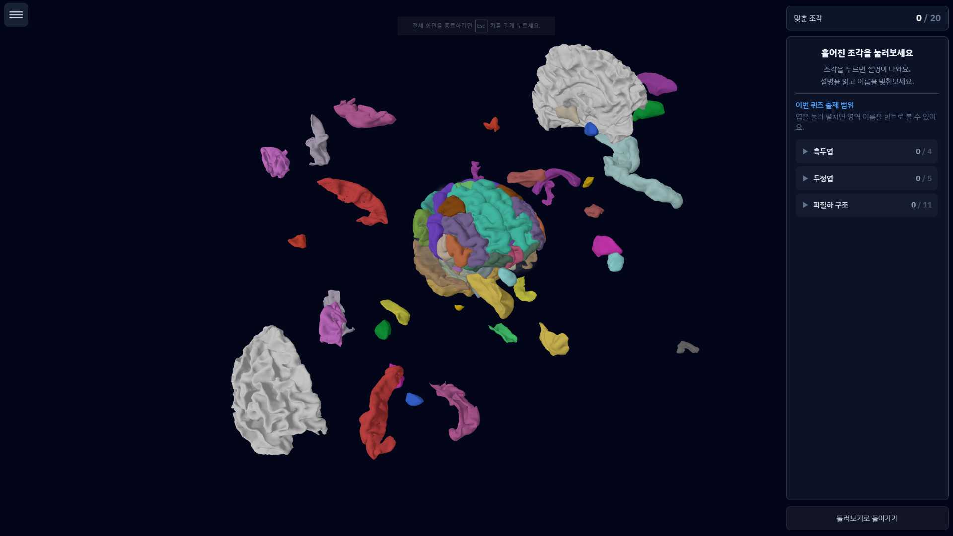
Spin it, explore it, then name it — a 3D brain plus quiz
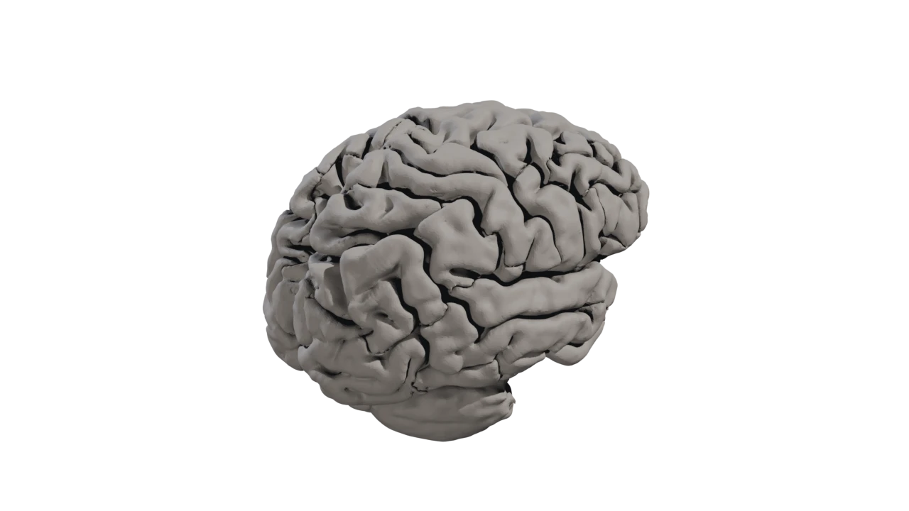
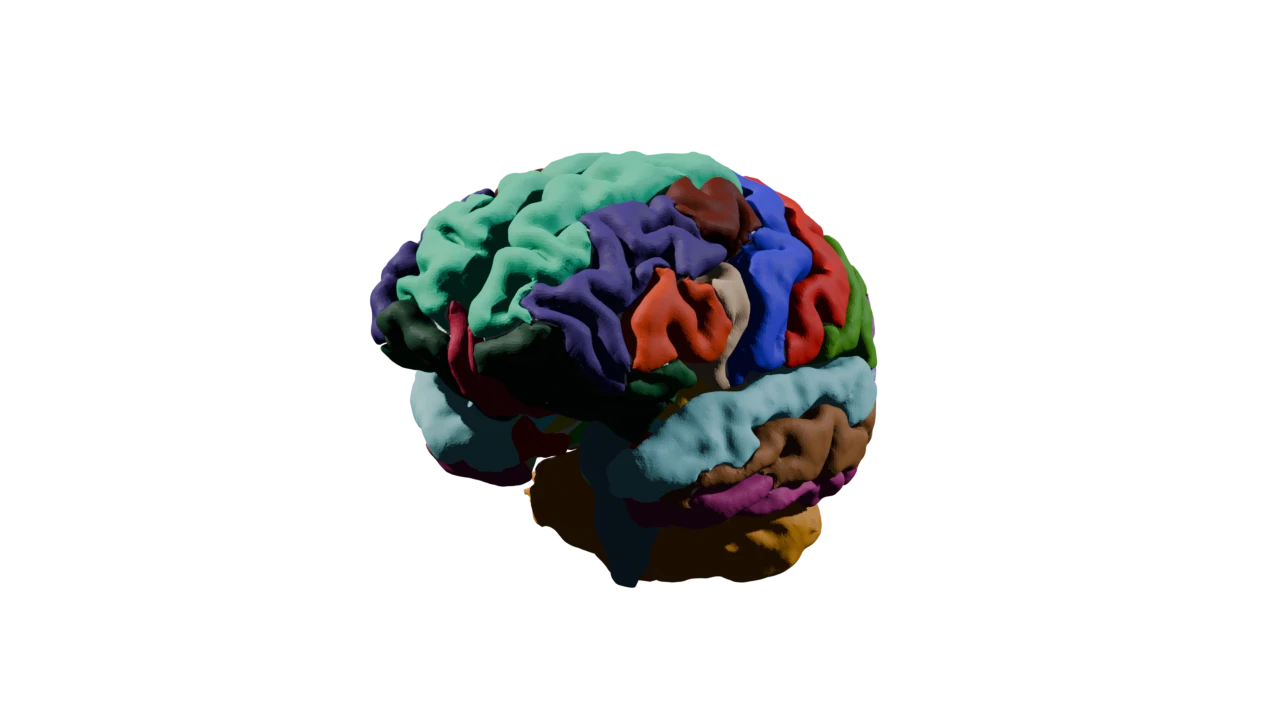
Explore the brain as if you were holding it. Spin and zoom a 3D brain that scatters into pieces and gathers back together, learn the names as you go, and lock them in with a name-the-region quiz. A section everyone enjoys, whatever their age.
03 / 05
04
CHAPTER 04 · How the brain ages
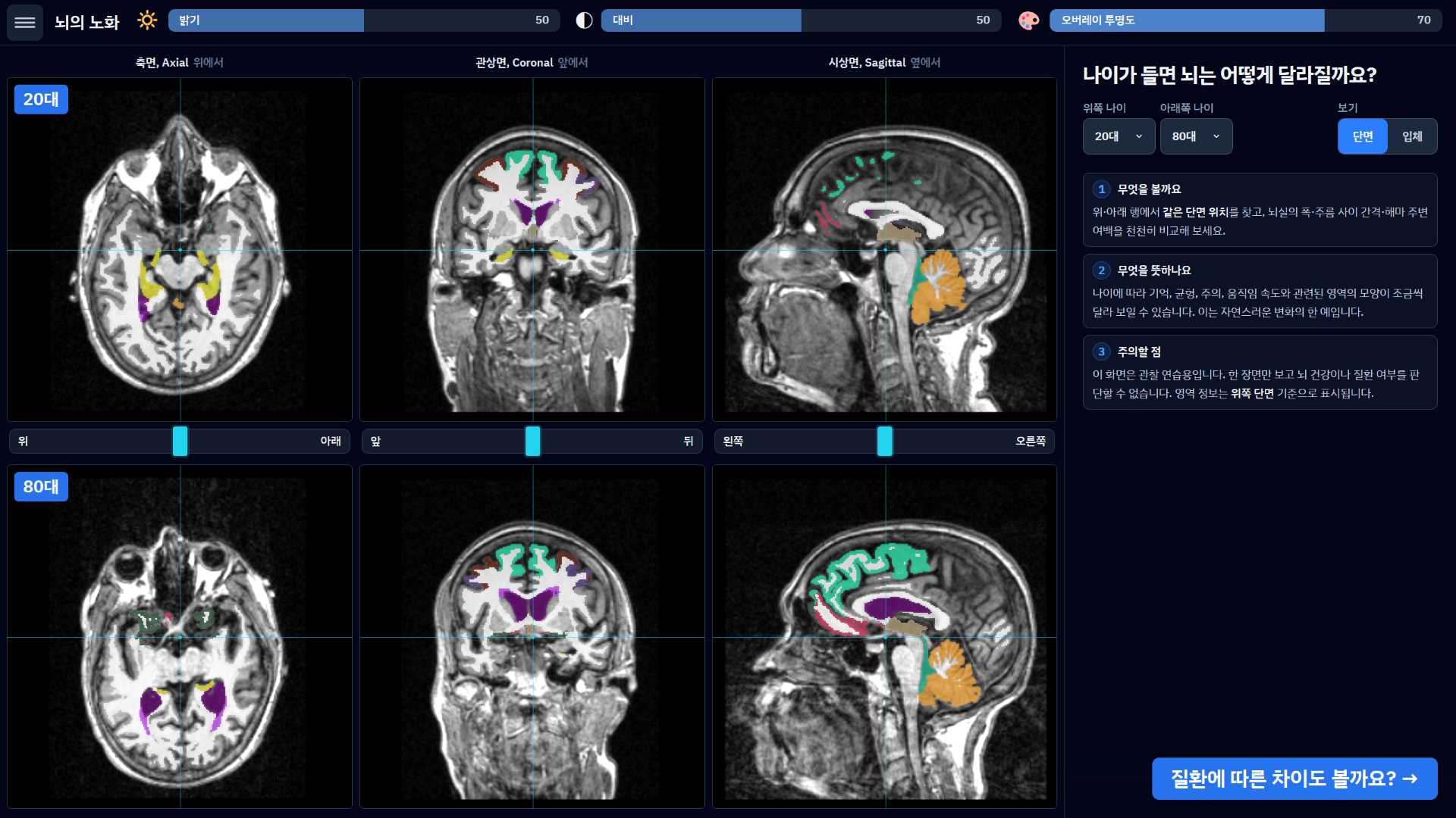
A young brain vs an older brain, side by side
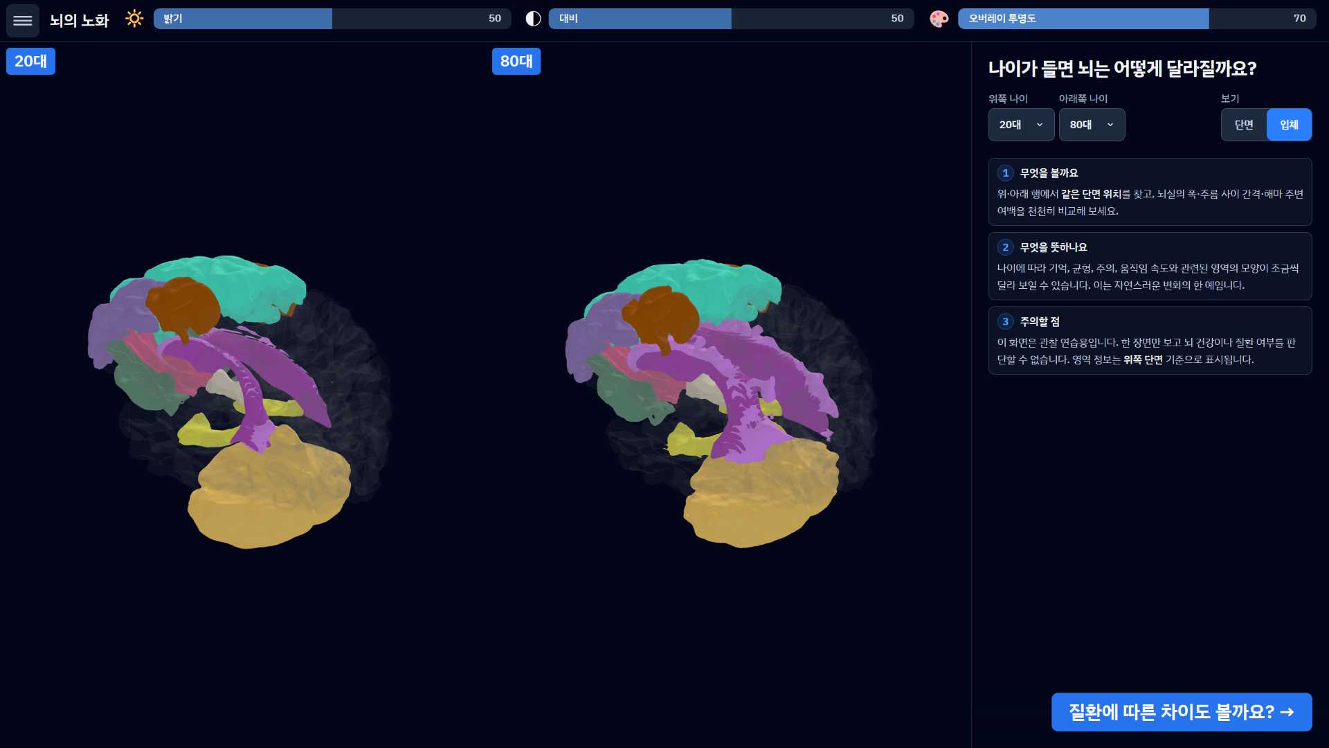
How does the brain change with age? Put MRIs of a young brain and an older brain side by side and compare. The regions that shift with age are picked out in color, so natural aging becomes something you can simply see.
04 / 05
05
CHAPTER 05 · Disease and how the brain changes
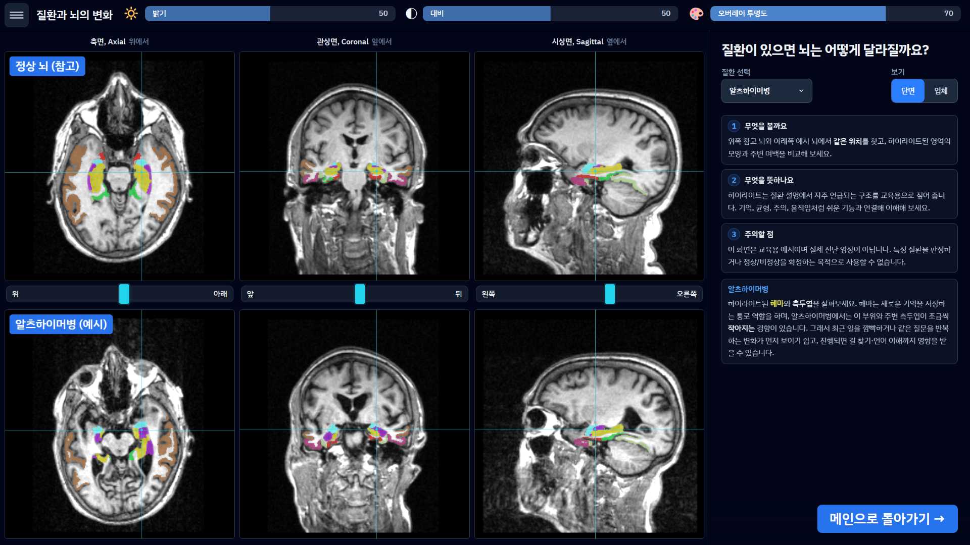
A healthy brain vs an affected brain, change by change
What is different between a healthy brain and one affected by disease? Compare two MRIs, one above the other, and look at the marks conditions such as dementia leave behind. Plain-language notes tell you which regions are involved.
05 / 05