MRI 영상 품질, 해결됐습니다.
인증된 고유전체 패드 + AI 분석. 임상 및 연구팀을 위해 설계되었습니다.
더 선명한 영상워크플로우 변경 불필요머리부터 발목까지AI + 하드웨어
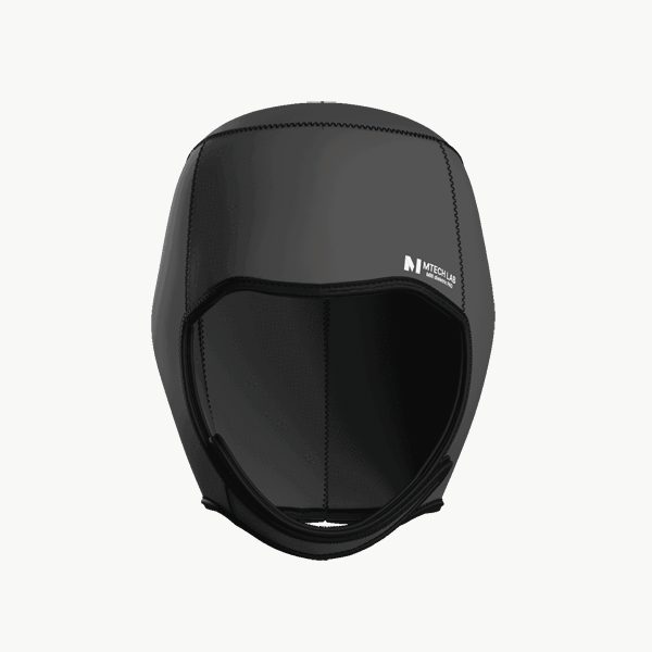
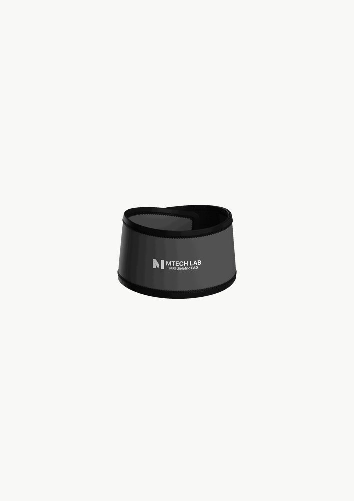
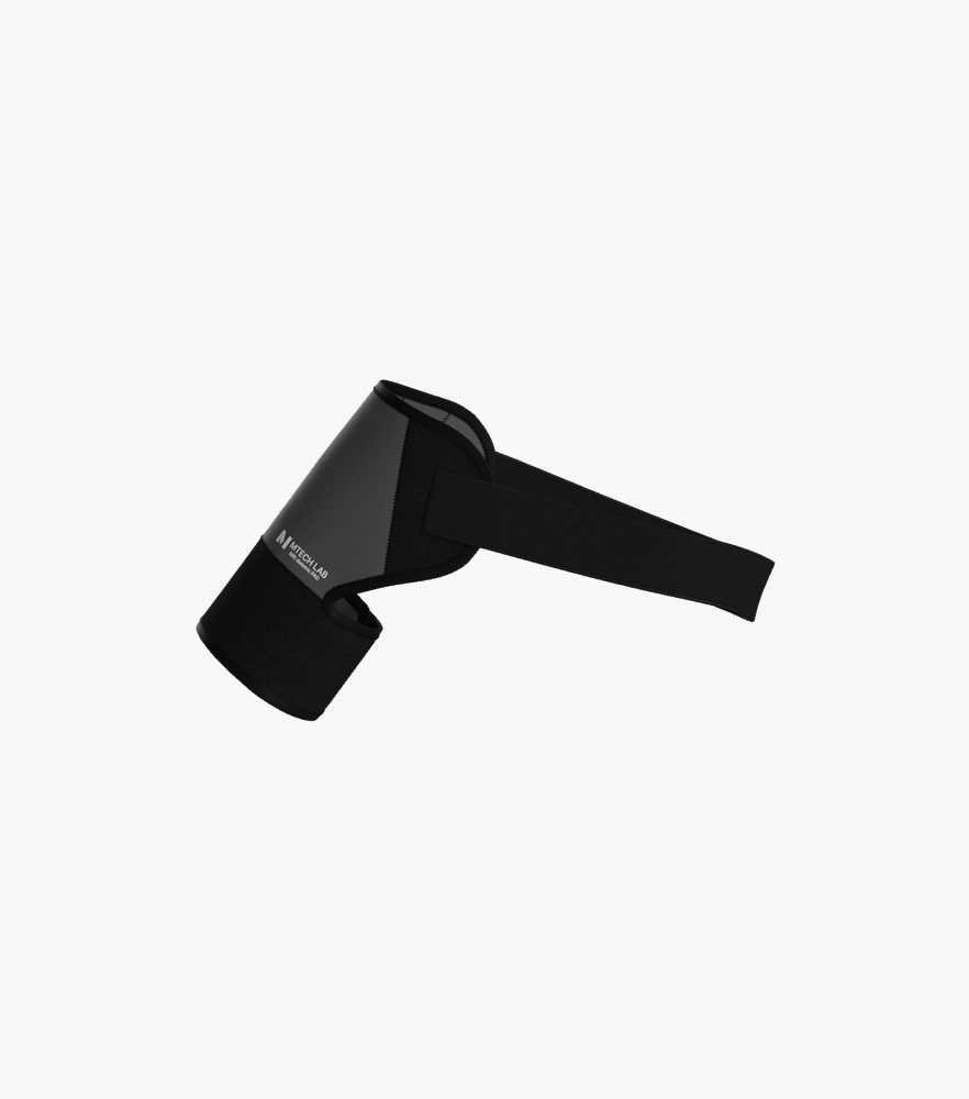
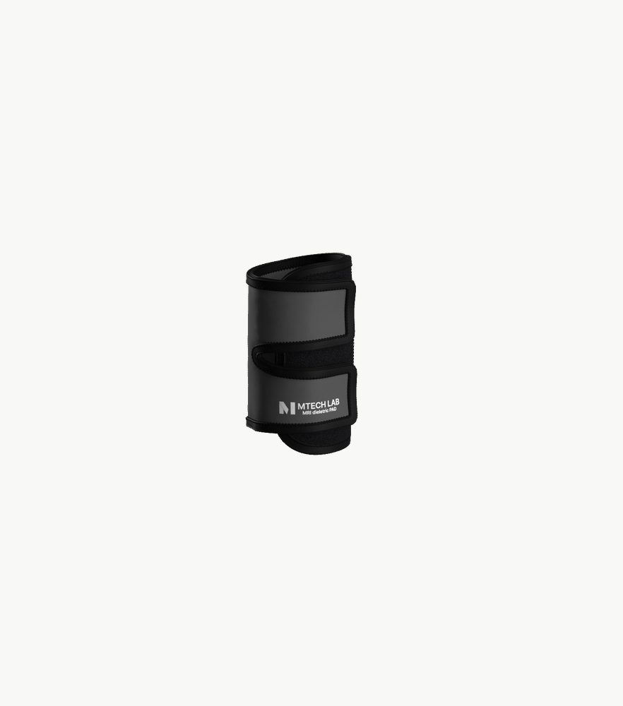
회사 소개 ›스캔하는 모든 해부 부위에 맞는 패드.
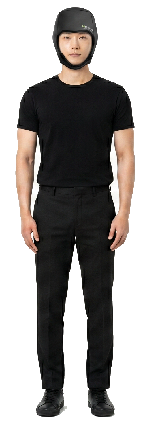
Male
Hover either figure to see pad details.
Features
- ✓High dielectric constant material contributes to B1 field homogenization
- ✓Provides clinically proven clear MRI brain images
- ✓Ergonomic design for better images
- ✓Harmless material to the human body
Fabric
- Outside
- Neoprene Polyester 100%
- Internal
- A high-gene substance
MRI 고유전체 패드


AI가 원본 영상을 통찰로 바꾸는 과정을 직접 확인하세요.
엠테크랩 AI 소프트웨어의 세 가지 가이드 영상 — 지능형 MRI 분석, PET 평가, 인터랙티브 뇌 해부 교육 — 으로 복잡한 영상 데이터를 명확하고 구조화된 임상 의사결정용 결과로 전환합니다.
탭을 선택하면 영상과 설명이 함께 바뀝니다.
AI MRI 분석 엔진
BRAINS — 지능형 MRI 분석 소프트웨어
MTechLab의 AI 분석 환경을 직접 들여다보세요. MRI 데이터 검토부터 자동 후처리, 그리고 원본 영상을 연구·임상에 바로 쓸 수 있는 명확한 인사이트로 전환하는 과정까지 — 하나의 통합 워크플로우로 이어집니다.
- MRI 검토부터 자동 처리까지 엔드투엔드
- 연구자와 임상의를 위한 구조화된 의사결정용 결과
- 실제 임상 영상 워크플로우 기반 설계