
Network connectivity
Region-to-region connectivity within large-scale brain networks from resting-state fMRI.

BrainDS + fMRI · Function
Research module · In development
BrainDS + fMRI is a research module in development. It will extend the BrainDS suite from structure and signal to function — how brain regions interact over time — for research hospitals, institutes and neuroscience teams.
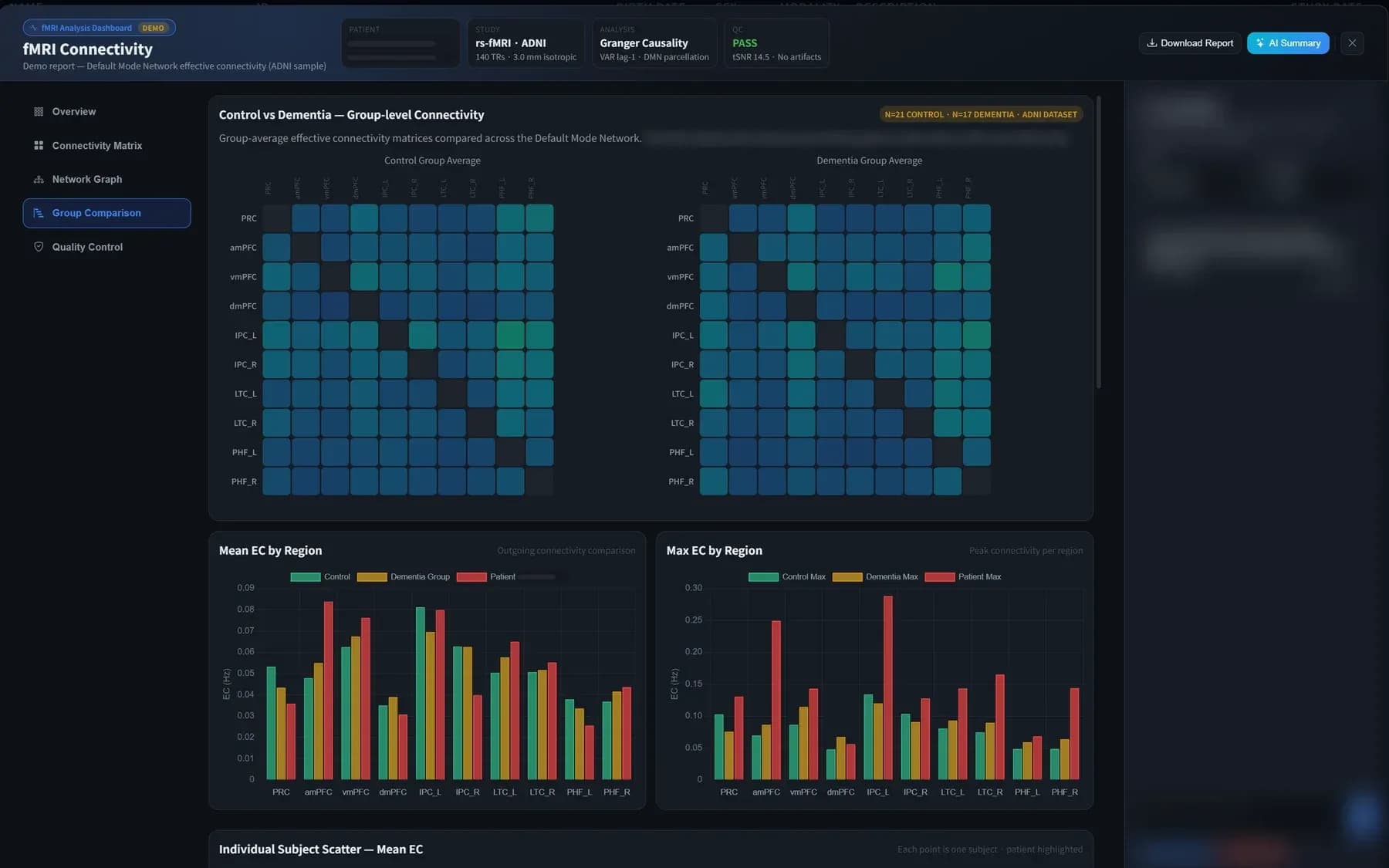
BrainDS + fMRI research prototype on demo data (ADNI sample), with a page from an internal rs-fMRI signal-quality report. Illustrative only; subject details masked.
Research module · In developmentNot yet available. Open to research collaboration; not intended for clinical use.
Volumetry and PET describe the brain at a single moment. Functional MRI records the BOLD signal over several minutes, so it can show which regions fluctuate together — a view of network organisation that neither structure nor tracer uptake provides.
Connectivity analysis treats the brain as a network: regions are nodes, and the strength of their co-fluctuation over time forms the links between them.
The planned workflow follows the same pattern as the rest of the suite. Every step below is in development.
01
Load a resting-state BOLD fMRI series together with the participant’s T1-weighted anatomical scan.
02
Report head motion and signal-quality measures first, so unreliable runs are identified before analysis.
03
Estimate connectivity between the regions of large-scale networks, such as the default mode network.
04
Compare individuals or groups with reference cohorts and export the results for research review.
Planned capabilities, shaped with research partners.
Region-to-region connectivity within large-scale brain networks from resting-state fMRI.
Connections explored as a matrix or an interactive graph, with thresholds to focus on the strongest links.
Individuals or groups compared with reference cohorts to highlight connections that differ.
Motion and signal-quality measures reported alongside every result.
Task-related activation maps for study designs that use tasks, complementing connectivity.
Designed to run in the same on-premise environment as BrainDS and BrainDS + PET.
Three views from the research prototype running on public research data: the network overview, a directed network graph and a region-to-region connectivity matrix.
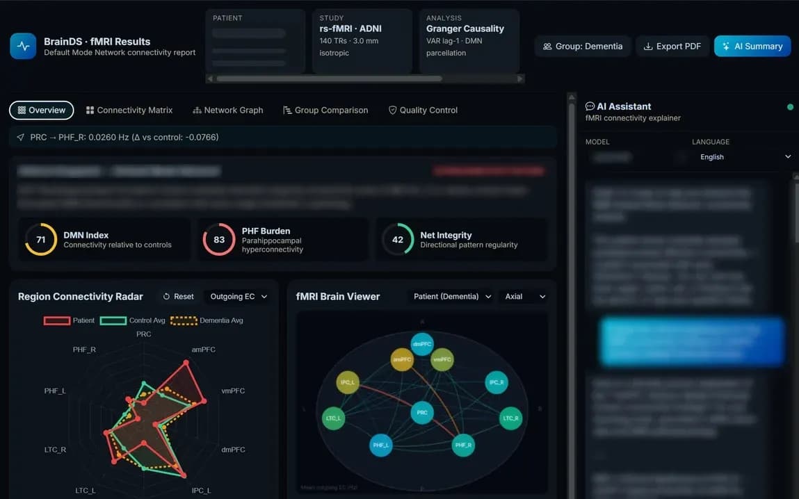
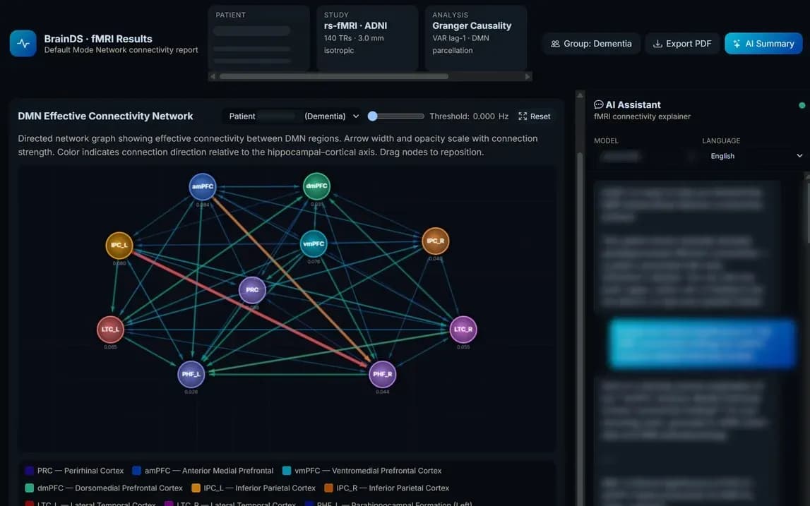
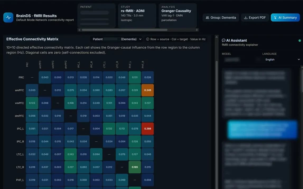
Research prototype — illustrative only. It is not part of the current BrainDS build, and the values shown are not validated results. Subject details are masked; assistant and summary text is blurred.
Connectivity estimates depend on a stable signal over time. In an internal single-volunteer pilot at 3T, median temporal SNR was 16% higher with MTechLab’s dielectric pad, with the largest gain in mid-brain slices.
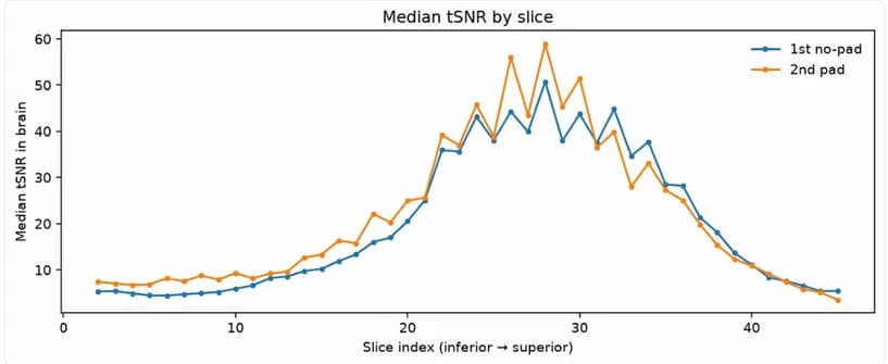
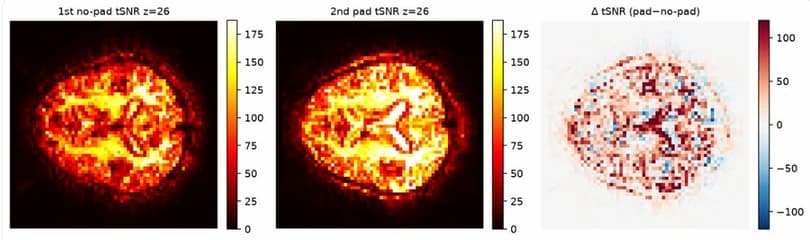
Internal pilot with one volunteer; the pad scan was acquired second and the data were not preprocessed. Not a validation study.
Members of our research team have published seed-based resting-state functional-connectivity studies at 3T. BrainDS + fMRI builds on that methodological experience.12
These are academic studies, not validations of BrainDS + fMRI.
References
Not yet. It is a research module in development. We welcome research partners who want to help shape its analyses and their validation.
Resting-state BOLD fMRI time series in DICOM or NIfTI format, together with a T1-weighted anatomical scan.
No. BrainDS + fMRI is intended for research use only.
It is planned as the third module of the suite: BrainDS measures structure, BrainDS + PET quantifies tracer signal, and BrainDS + fMRI will analyse function.
We are looking for research hospitals, institutes and neuroscience teams to collaborate on BrainDS + fMRI.